Specimens of the Department of Botany
Vascular Plant
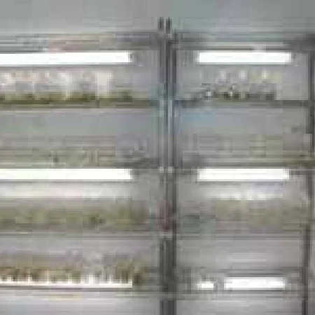
The vascular plant specimen storage, located on the 2nd and 3rd floors of the Department of Botany Building and the 5th floor of the Natural History Collection Wing in the Tsukuba District, currently houses approximately 1.2 million pressed specimens. In 1925, Tomitaro Makino and Kanji Nemoto published "Flora of Japan," which was based on specimens collected to elucidate the flora of Japan since the Museum's establishment in 1877. This book became a standard reference for Japanese botanists at the time.
A field guide to wild plants was published by Shunki Okuyama, an Honorary Member of the Museum, based on specimens donated by plant enthusiasts from across Japan for the pressed flower exhibitions held from 1933 to 1970. This publication made a significant
contribution to educational outreach activities. The Museum's collection has been expanded with specimens from Europe and America acquired through exsiccatae exchange, as well as with those collected by Museum staff members such as Takenoshin Nakai, Yoshisuke
Satake, Shunki Okuyama, and Jisaburo Ohwi. Building on this, the late Dr. Jisaburo Ohwi, an Honorary Member of the Museum, published, "Flora of Japan - Spermatophytes" in 1953, a comprehensive work on Japanese seed plants. The English version was published
in 1965. Today, it remains an indispensable reference for compiling regional floras. The Museum continues to exchange exsiccatae with international partners, collecting specimens from regions such as North America and various Asian countries.
More recent additions to the collection include specimens gathered by current staff members during surveys in Japan and abroad. Domestically, as part of an integrated survey of the Japanese archipelago, specimens have been collected all across the country,
from Hokkaido in the north to Okinawa in the south. Abroad, specimens have been collected through joint surveys in locations such as the Himalayas, Thailand, New Guinea, China, Siberia, Vanuatu, and Myanmar. Additionally, a collection of cherry species
native to Japan, including horticultural varieties, has been compiled by Tetsuya Kawasaki.
To date, approximately 2,700 type specimens, which serve as the reference for scientific names, have been identified. The historical collection includes about 1,400 specimens that served as the basis for "Somoku-zusetsu" (Figures and Descriptions of Plants) by the late-Edo period botanist Yokusai Iinuma, about 300 specimens collected by botanist Keisuke Ito and his grandson Tokutaro from the late Edo to the early Meiji period, and about 500 specimens collected in Tibet by Ekai Kawaguchi, who visited the region from the late Meiji to the Taisho period.
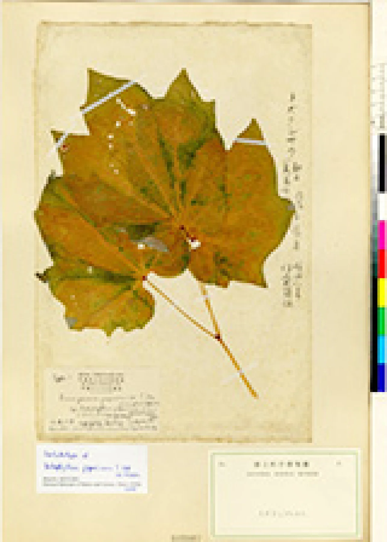 One of the type specimens of Ranzania japonica, described by Tokutaro Ito, the first Japanese botanist to assign a scientific name to a plant
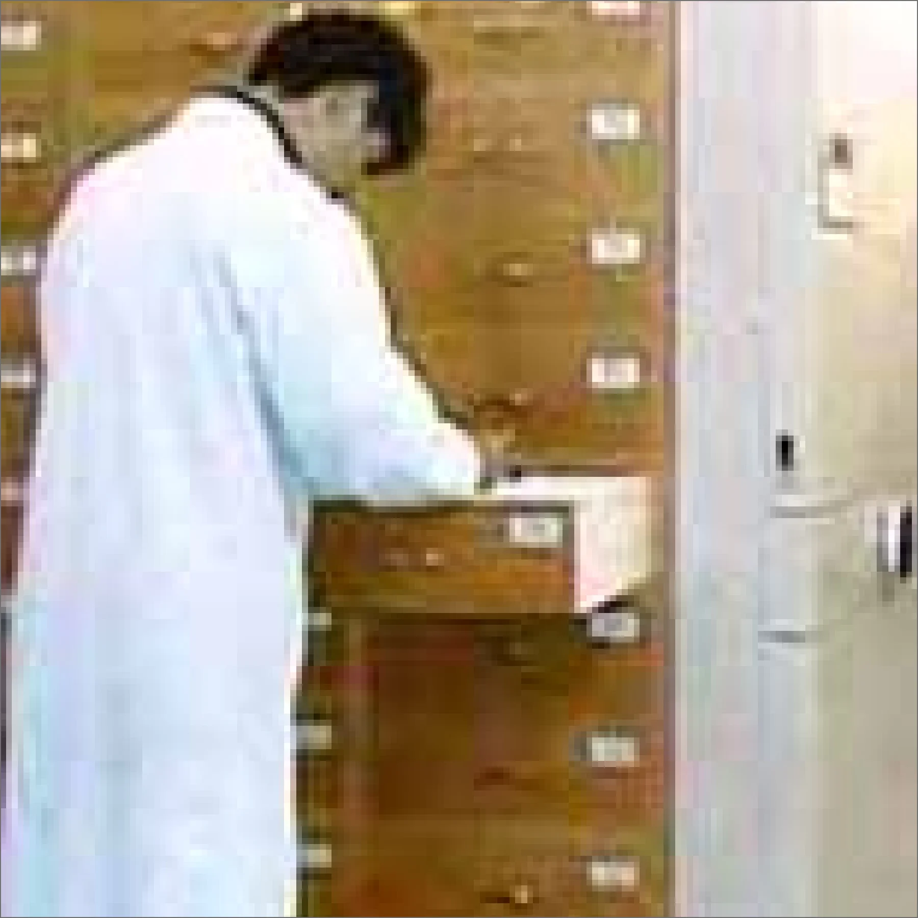
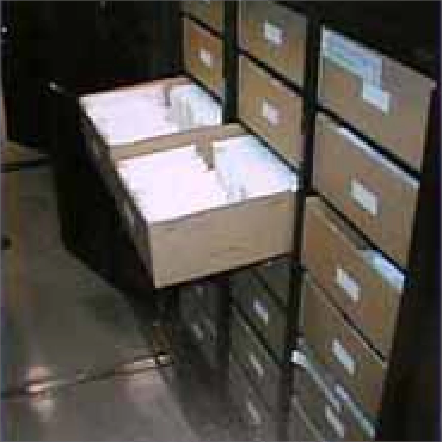
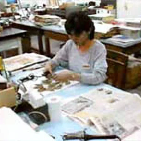 Specimen preparation (mounting process)
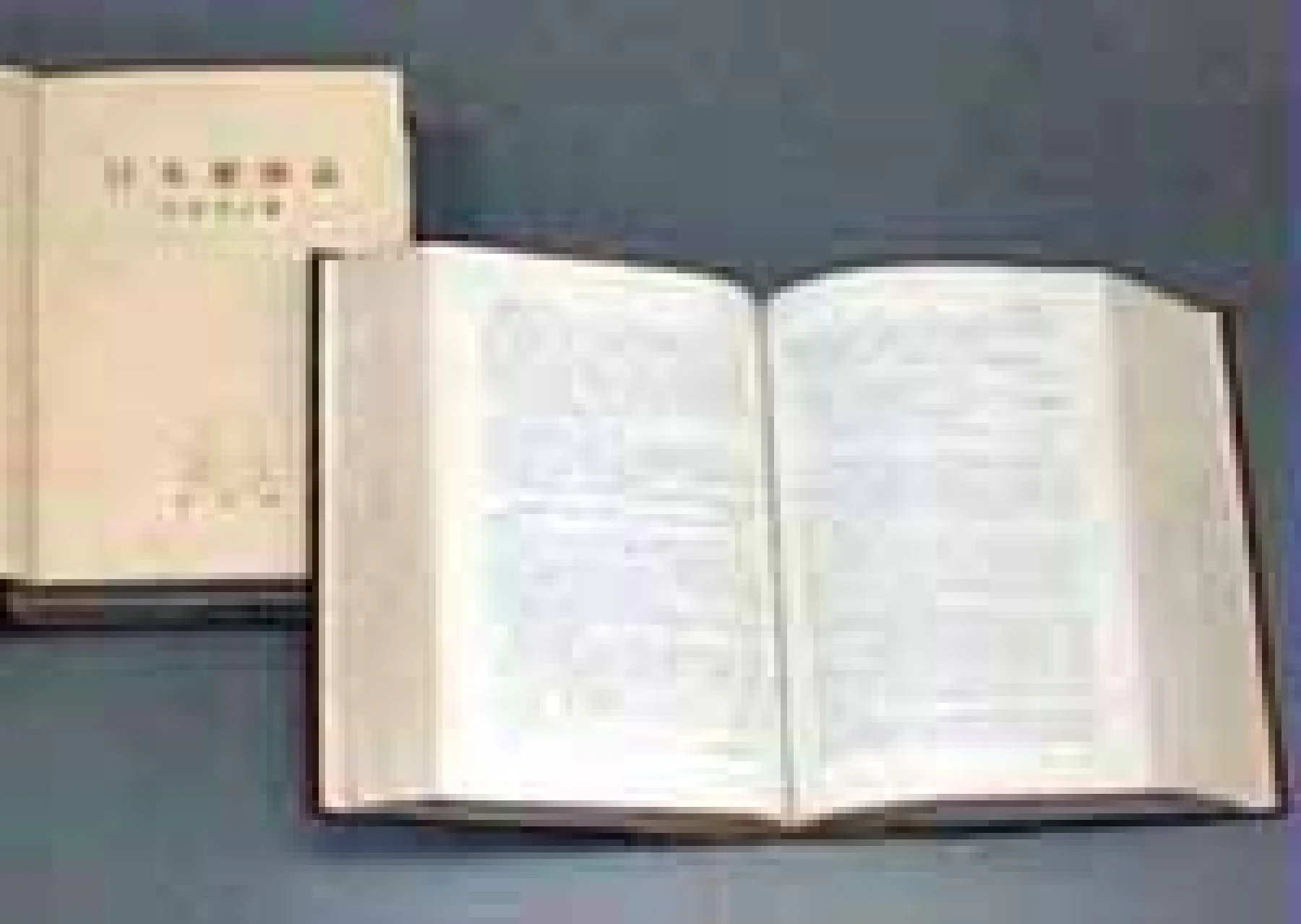 "Flora of Japan - Spermatophytes" by Jisaburo Ohwi
Bryophytes
The Bryophyte Collection Room houses about 240,000 specimens.
These specimens consist primarily of items gathered from Japan and abroad by our staff members—Sinsuke Hattori (in office 1941–45), Hiroshi Inoue (1962–89), Masanobu Higuchi (1993–2021), and Yuya Inoue (2021–present)—through collection and exchange, as
well as donations to the Museum.
Notable specimens include:
(1) Type specimens,
(2) Individual collections of Hisahiko Sasaoka, Hiroshi Inoue, Takemasa Osada, Kamezo Saito, Ryozo Watanabe, Iwao Nagano, etc.,
(3) Taxonomic group collections, including the genus Plagiochila from Hiroshi Inoue and the family Pottiaceae from Kamezo Saito.
and (4) Regional collections from New Guinea, South America (Chile, Peru), and the Himalayas (Nepal, Pakistan).
For easy access, basic information for about 1,150 type specimens—including species names, published literature, publication years, and collection localities—has been compiled into a database that is searchable on the National Museum of Nature and Science website.
The Hisahiko Sasaoka collection consists of about 10,000 moss specimens (including about 120 type specimens) collected in Japan in the early 1900s, and these have played an important role in elucidating Japan's moss flora. Assembled from around the world by Hiroshi Inoue, an expert on the genus Plagiochila, this collection of about 5,000 specimens constitutes the world's most comprehensive collection of this genus in terms of both quality and quantity. The Museum's regional collections from New Guinea, South America, and the Himalayas were collected through overseas surveys funded by Grants-in-Aid for Scientific Research, and specimen collection is still ongoing, primarily in Asia and Oceania. These specimens provide the foundation for research elucidating the bryophyte flora of these regions and for taxonomic studies of various groups.
Bryophytes are generally smaller than vascular plants, so specimens collected in the field are typically not pressed. Instead, they are air-dried and stored in specimen bags (11 cm x 15 cm). A single folded sheet of paper is used for specimen bags worldwide, a design that allows the contents to be easily viewed when unfolded. The specimens are arranged upright like cards in the drawers of wooden cabinets and arranged alphabetically by scientific name within the three groups of bryophytes: mosses, liverworts, and hornworts. Larger specimens are placed in specimen bags of an appropriate size or mounted on sheets. Type specimens, larger specimens, and specimens mounted on sheets are stored separately from the main collection.
 Specimens are rehydrated, prepared as slides, and then its various parts are examined under a microscope
 Bryophyte Collection Room

Macroalgae
Algae comprise many phylogenetically diverse groups and vary remarkably in size, ranging from micrometer-scale phytoplankton to kelp that can grow to tens of meters in length. Consequently, the morphology of these specimens is also highly varied. For practical purposes, our research department divides its collection into two management categories: microscopic-sized microalgae (primarily unicellular) and macroscopic-sized macroalgae (multicellular). For the macroalgae featured here, the primary preservation methods are pressed specimens and liquid-preserved specimens.
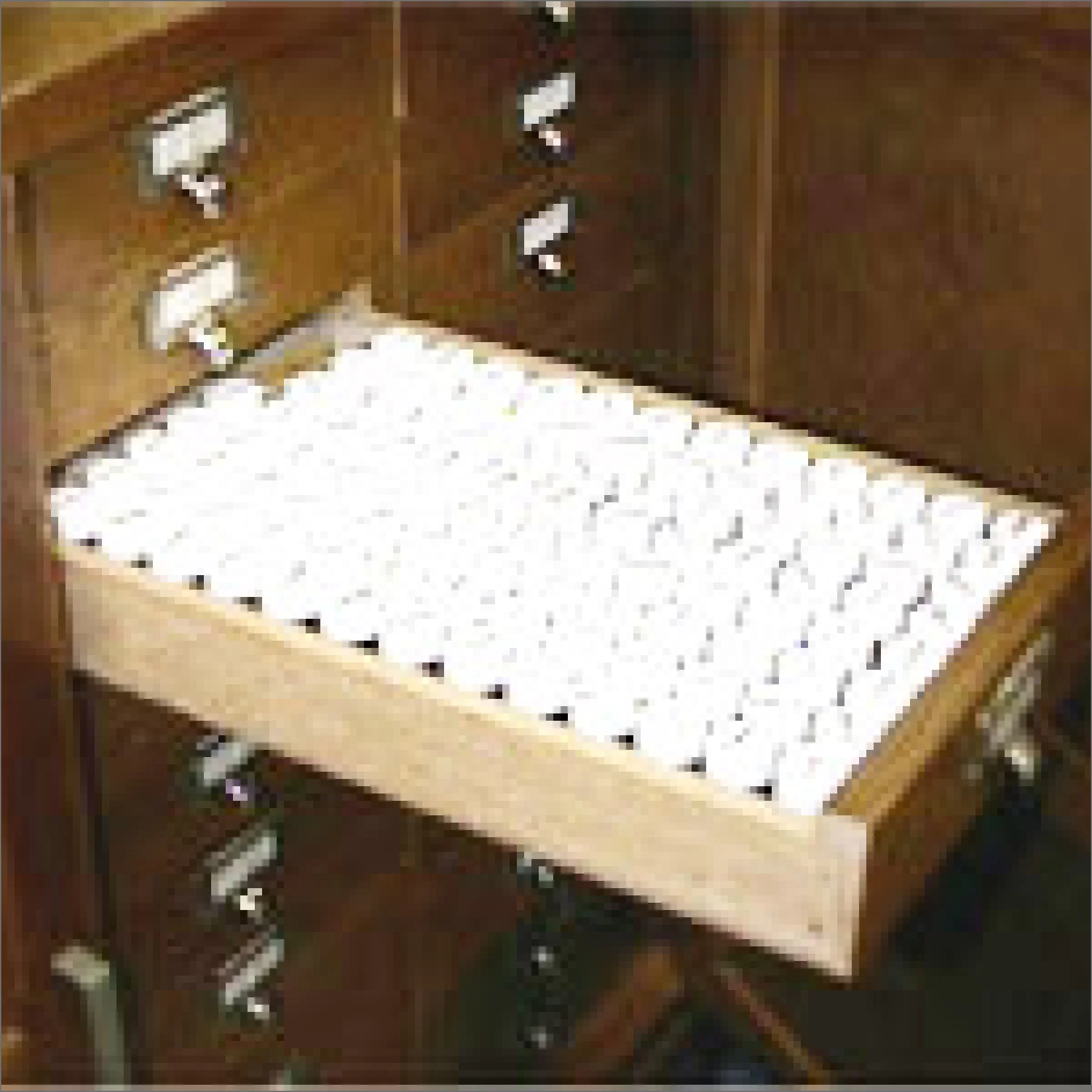
Leaf Specimen Collection Room
The Lead Specimen Collection Room houses approximately 65,000 items, consisting of specimens collected throughout Japan and exsiccatae donated by overseas institutions. Like vascular plants, these specimens are dried and mounted on sheets for preservation.
Most macroalgae can be classified as green, brown, or red algae, so in this herbarium, the general specimens are divided into these three categories and then arranged alphabetically by their scientific names. A database of the collection is currently
under development, allowing users to search for green and brown algae.
Special specimens are stored separately from the general collection. These include type specimens (about 90 items), specimens identified by Kintaro Okamura (about 1,400 items), and the Michitaro Higashi Collection. Kintaro Okamura (1867–1935), a renowned
pioneer of Japanese phycology, made numerous contributions to the study of seaweeds along the Japanese coast from the Meiji to the early Showa period. A database of the specimens identified by Okamura is now complete, enabling it to be searched on a computer.
One of his most important works is the "Icones of Japanese Algae," published in seven volumes from 1907 to 1942. This herbarium holds some of the original illustrations for the first four volumes.
Liquid-Preserved Specimen Collection Room
Seaweeds are often preserved in fixative solutions because they tend to lose their original shape when dried. This collection room houses about 10,000 liquid-preserved specimens. Additionally, specimens of giant algae such as giant kelp are preserved
through glycerin impregnation. The specimens are arranged in order of registration, with approximately 2,000 of them searchable using both cards and computers.
 Specimen of Dudresnaya japonica Okamura, identified by Dr. Kintaro Okamura. Collected by Kotaro Saita in 1894.
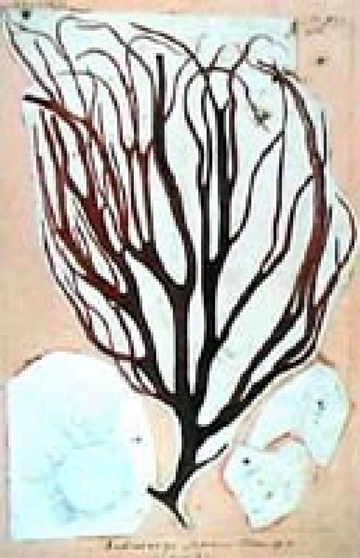 Original illustration used by Dr. Kintaro Okamura in 1908 for the original description of Dudresnaya japonica Okamura as a new species.
Microalgae
Many freshwater algae are microscopic, and in their natural habitat, they typically grow in mixed communities with numerous other species. Since it is impossible to collect these algae individually or by species in the field, they are gathered in mixed
samples containing hundreds or thousands of individual specimens. These samples are then placed in 20 ml specimen bottles and preserved with formalin. The specimen bottles are labeled with serial numbers so they can be cross-referenced with the collection
records. They are stored in a dedicated cabinet in the liquid-preserved specimen collection room. In addition to these liquid-preserved specimens, other materials are also stored as specimens, including permanent slides prepared from them and items for
electron microscopy such as membrane filters, SEM stubs, and TEM blocks. In the study of microalgae, sketches and photographs may also be treated as specimens, so original illustrations, photographic plates, films, and slides of the specimens are also
carefully preserved.
As of the end of fiscal year 2020, our collection totals 44,000 items. This includes a general collection from the Museum's own research activities—notably specimens from the Himalayas and Papua New Guinea collected by Mr. Masayuki Watanabe (former Director
of the Department of Botany)—as well as donated collections from Haruo Okuno, Toshiharu Watanabe, Minoru Hirano, Hiroshi Fukushima, Takaaki Yamagishi, and Makoto Mizuno.
Freshwater algae are small organisms, and many groups are difficult to classify due to a lack of distinct taxonomic features. Just as the classification and identification of seed plants often requires observing their flowers, it is often impossible to
identify microalgae—which have few distinct morphological features of the vegetative body—even at the genus level without observing the shape of their reproductive cells and their methods of reproduction. To facilitate this, the Department of Botany Building
is equipped with a culture room for isolating and culturing samples collected in the field, allowing for continuous observation of their life cycles. It also has an algal strain preservation room to support various research methods, including genetic
analysis.
 Liquid-preserved specimens
 Algal strain preservation room
Lichens
Lichens (リンクを新しいタブで開きます)are symbiotic organisms of fungi and algae found on rocks, soil, and tree bark. The thallus (the body of a lichen) varies in size, ranging from under a few millimeters in diameter to over several meters. Their growth forms are also incredibly diverse, ranging from those that adhere tightly to rocks and tree bark to more complex, leaf-like or branched structures.
The classification of lichen species is based not only on the morphological characteristics of the thallus and reproductive organs, but also on the types of chemical components within the thallus, known as lichen products. Lichens collected in the field are carefully arranged and stored on specimen shelves to facilitate observation of their morphology and testing for lichen products. Consequently, lichen specimens come in various forms. These include specimens pressed like flowering plants, rocks with attached crustose lichens mounted directly onto sheets, and others kept in small boxes.
The Department of Botany houses a collection of about 250,000 lichen specimens gathered from a wide range of regions across the globe, including the Antarctic, Arctic, and tropics. These are organized into general specimens (about 230,000), exsiccatae specimens (about 20,000), and type specimens (リンクを新しいタブで開きます)(about 1,000). The general and type specimens are arranged alphabetically by genus and specific epithet, and then grouped by locality. "Exsiccata" is a Latin term for "dried plants" that has come to mean a collection of dried plant specimens. These collections contain numerous isotype specimens and are extremely valuable for advancing taxonomic research. The National Museum of Nature and Science also publishes the lichen exsiccata "Rare Lichen Specimen Collection(リンクを新しいタブで開きます)" (1994-2008, 2010-).
The lichen collection at the National Museum of Nature and Science, initially based on a donation of approximately 80,000 specimens from Dr. Yasuhiko Asahina, has since been expanded and curated with specimens collected by Sho Kurokawa, Hiroyuki Kashiwadani, Yoshihito Ohmura, and others. This collection is recognized worldwide for its exceptional quality and quantity. It is utilized by researchers from Japan and abroad and is cited in research papers more than 100 times annually.
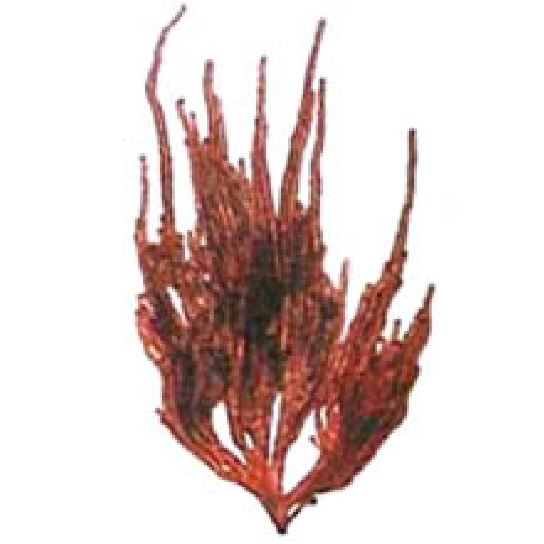
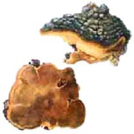
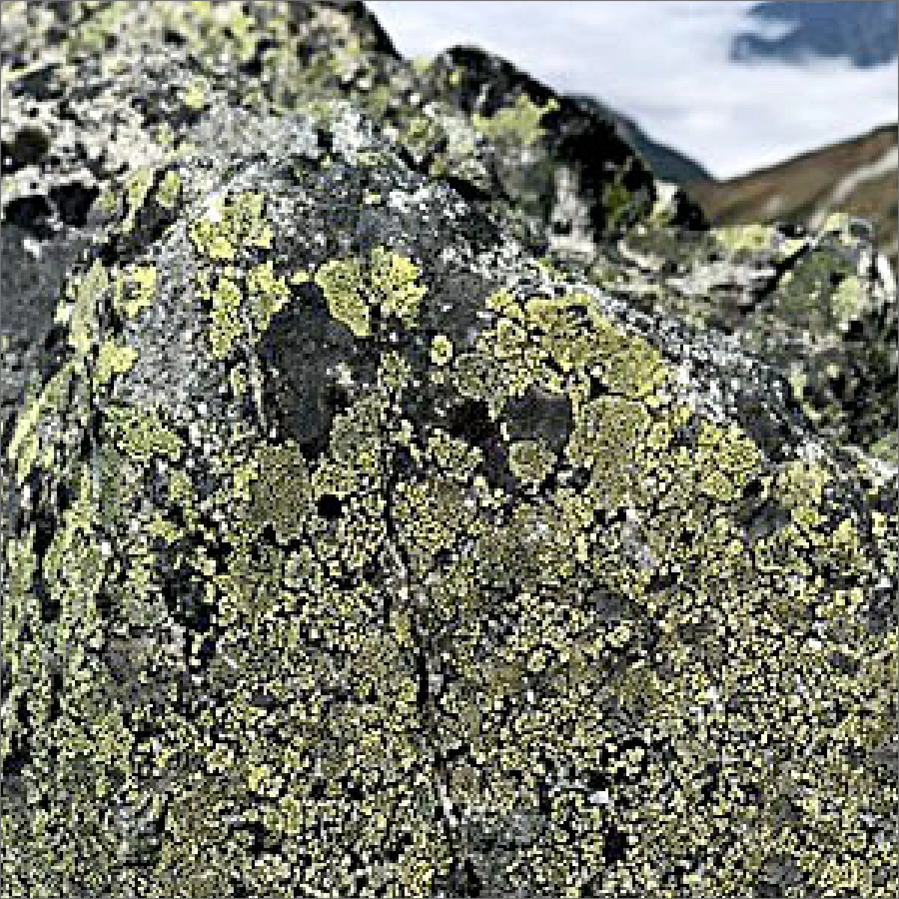 Growth of map lichen
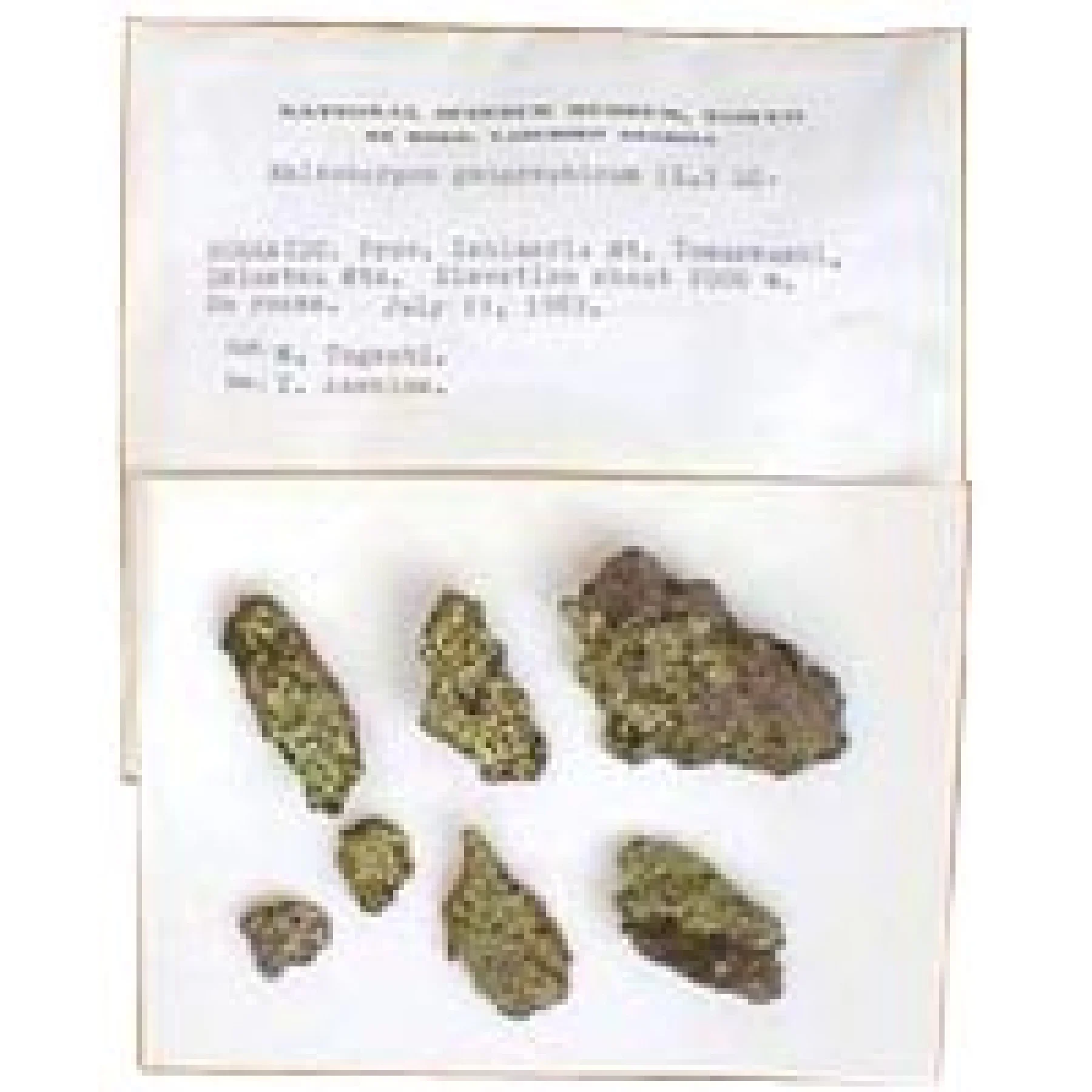 Specimens of crustose lichens are collected together with the rocks on which they grow, mounted on thick sheets, labeled, and then organized and stored in specimen bags.
Fungi
The specimen room on the third floor of the Department of Botany Building in the Tsukuba District houses dried specimens of chytrids, zygomycetes, ascomycetes, basidiomycetes, and imperfect fungi (according to the old classification). Additionally, the specimen room on the fifth floor houses oomycetes and hyphochytrids, which are now commonly referred to as pseudofungi. The total number of specimens is approximately 150,000. Most of these general specimens are stored in standard paper bags or boxes. In addition, there are specimens slides, specimens of large fungi, and liquid-preserved specimens (including type specimens of fungi such as caterpillar fungus). The Museum's collection of fungal specimens includes over 3,000 type specimens and other important collections.
Important specimen collections include those from numerous researchers, such as Atsushi Yasuda (primarily mushroom specimens from the pioneering era of Japanese mycological research), Kanesuke Hara (microfungi, including plant-pathogenic species), Mitsutaro Shirai and Shunsuke Kusano (microfungi, including plant-pathogenic species), Kaneyoshi Sawada (fungi native to Taiwan), Yoshikazu Nishikado (plant-pathogenic fungi and historical exsiccatae from 19th-century Europe), Kogo Togashi (primarily tree-pathogenic fungi), Yasu Homma (primarily Erysiphaceae), Rokuya Imazeki (mainly polypores), Yoshio Kobayashi and Daisuke Shimizu (caterpillar fungus, etc.), Tsuguo Hongo (Agaricales), Yoshio Otani (primarily Pezizales), Yukihiko Nomura (primarily Erysiphaceae), and Yoshimichi Doi (primarily Hypocreales). The collection also features specimens obtained during surveys of endangered species, conducted with the cooperation of citizen scientists from various regions. In addition to domestic specimens, the collection includes foreign fungal specimens collected by museum staff during overseas surveys in regions such as Papua New Guinea, Alaska, South America, New Zealand, Tasmania, New Caledonia, China, and Nepal, as well as exchanged specimens (exsiccatae). These specimens are invaluable for comparative research on the taxonomy of fungi native to Japan.
 Arrangement of bagged specimens
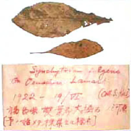 The first Synchytrium specimen from Shunsuke Kusano's research, which was instrumental in establishing Japanese mycology on the world stage
 Hypocrea splendens, listed as an endangered species in the Red Data Book.
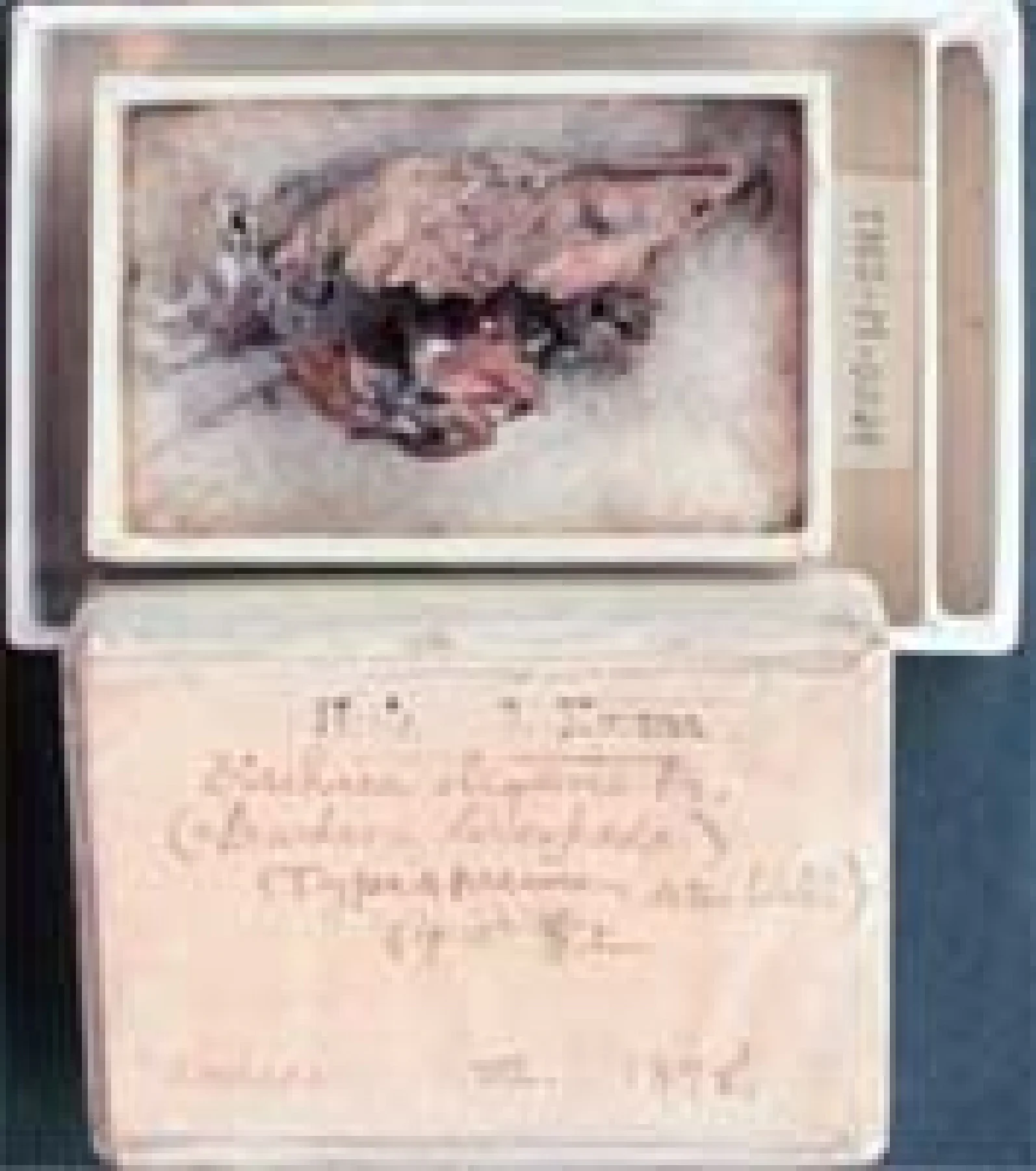
 Seiichi Kawamura's original illustration, "Phellinus linteus"
Slime Molds
As of March 2018, the collection houses about 74,000 specimens. These include four major collections of great value not only for scientific research but also for the history of Japanese slime mold taxonomy.
Kusano Collection (about 200 specimens)
These slime mold specimens were collected by Mr. Shunsuke Kusano (1874-1962) from the Koishikawa Botanical Garden and Komaba Farm while he was studying and working at the Tokyo Imperial University. These are the first Japanese slime mold specimens to
be formally collected by a Japanese individual, and they include duplicates of specimens cited in the first report on Japanese slime molds, published by A. Lister in England in 1904.
Minakata Collection (about 6,000 specimens)
This historically valuable collection of slime mold specimens was amassed by Mr. Kumagusu Minakata (1867-1941), a pioneering figure in the study of Japanese slime mold taxonomy. The collection also includes many specimens collected by his students, as
well as those sent from across Japan for identification. Additionally, the Museum houses thousands of illustrations of mushrooms and molds known as the Minakata Kumagusu Colored Illustrations, which consist of a set of illustrations, specimens, and description
Koaze Collection (about 16,000 specimens)
This collection of slime mold specimens was assembled by Mr. Shiro Koaze (1875-1951), a student of Kumagusu Minakata. The specimens were collected from across Japan to aid in the study of morphological variation among species. Containing over 100 unpublished
new species, the collection is vital for elucidating the slime mold flora of Japan, and some of its specimens have already been cited in the "Illustrated Guide to Japanese Slime Molds" by Yukinori Yamamoto, published in 1998.
Emoto Collection (about 2,000 specimens)
This collection of slime mold specimens was amassed by Mr. Yoshikazu Emoto (1892-1979). Its core consists of specimens cited in "Nova Flora Japonica No. 8, Slime Molds," the first monograph on slime molds in Japan, which was authored by Emoto and published
in 1942. Its original color illustrations were not published until 35 years later, in 1977. It includes 10 type specimens. Another key feature is the inclusion of numerous specimens from overseas exchanges, representing more than 200 species.
In addition to these collections, the Museum is currently in the process of accessioning about 25,000 slime mold specimens collected by Yukinori Yamamoto (1947–), who remains active in the field. Furthermore, the number of slime mold specimens accepted from the Japanese Society of Myxomycetology (founded in 1977) has surpassed 12,000, and the Museum continues to accept more. The specimens donated by the society significantly contribute not only to research but also to the Museum's exhibitions and educational outreach activities. In addition, the Museum's collection of slime mold specimens from the Himalayan region and other areas, resulting from its own overseas survey expeditions, now exceeds 1,000. All of these specimens have been cited in various publications, including a series of research papers on Himalayan slime molds.

 The oldest slime mold specimen collected by a Japanese individual
 Original color illustrations from the Emoto Collection
