
Extraction of Beaks from Buccal Mass
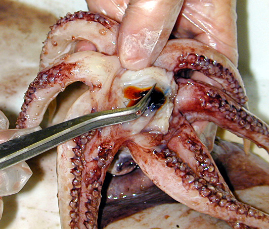 Pick up tip of rostrum with forceps 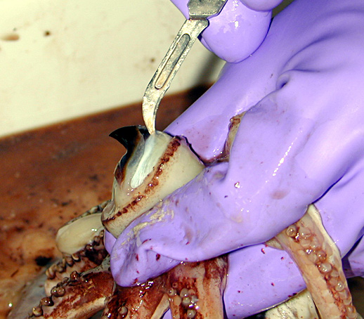 Cutting the muscle from the crest 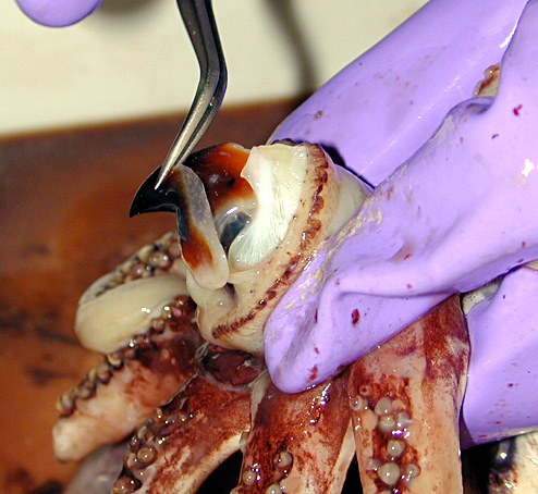 Severe the muscle from the wing and pull out the beak Severe the muscle from the wing and pull out the beak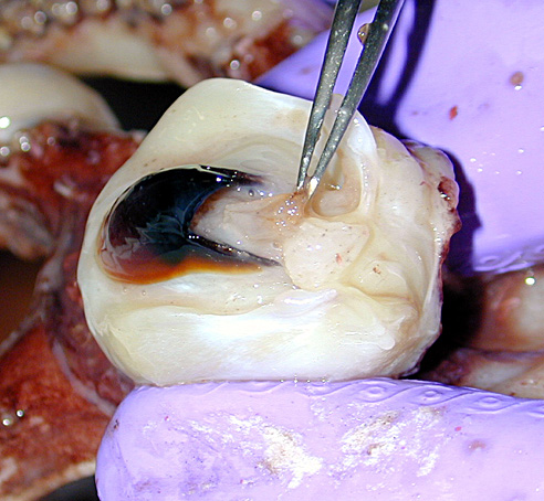 Remove the radula 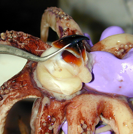 Remove the upper beak |
It is necessary to remove beaks from the
buccal mass of specimens in digestive stages 3-4; however it is very difficult
to remove beaks after the specimen has been preserved in formalin because of
shrinkage of the buccal mass muscle. On
such occasions if you are forced to remove the beaks, wing and lateral walls
are easily damaged. Therefore use the
following methods for removal of beaks to avoid damaging them:
1. If the buccal mass is still in the arm-head part it will first need to be removed. It is relatively easy to remove the buccal mass by cutting between the 4th arm pair. Attachment of buccal membrane to the arm is an important systematic character so pay attention not to damage the buccal membrane. Posterior to the buccal mass is the esophagus, which needs to be cut with scissors to avoid damaging the buccal mass. 2. The buccal mass is a round, muscular ball. The tips of the upper and lower beaks and the wings of the lower beak are exposed. Pick up tip of lower beak and pull posteriorly, gently. Pay attention not to damage the wings and lateral walls. After removing the lower beak, remove the upper beak using the same method. When removing the upper beak you will see a tongue-like structure with the radula on it. It is better to remove the radula with forceps. Place the upper and lower beaks, along with radula, muscle tissues, and head-arm parts in a jar of alcohol with a label. 3. When it is difficult to remove the lower beak, soak the buccal mass in 1-3% of NaOH or KOH for several hours. But as Clark (1986) pointed out, if soaked for too long the cartilaginous and wing parts can be dissolved, so remove the buccal mass when it becomes soft and rinse with water. 4. Even after soaking it is sometimes difficult to pull out the beaks. In this case, you can slide a thin metal plate (not too sharp) between the wing and muscle of the buccal mass and severe the connection between the two. Do this on both sides. Next use a scalpel to cut the muscle from the crest. Then place the thin metal plate between the lateral wall and the muscle to separate the two. Then pull out the beak. The upper beak is removed in the same way. Sometimes it is difficult to remove the crest from the muscle so be careful not to break the beak. In this manual lower beaks are mainly used for identification so upper beaks can be left in the buccal mass when it is difficult to remove them. Using the methods outlined above beaks can be removed from specimens identified with external morphology or ordinary systematics. It would be useful to make a reference collection of beaks removed from these accurately identified species. Click on an image to see an enlarged view; click X to close. |
All Rights Reserved Kubodera, 2000